Institute of Molecular Genetics of the Czech Academy of Sciences
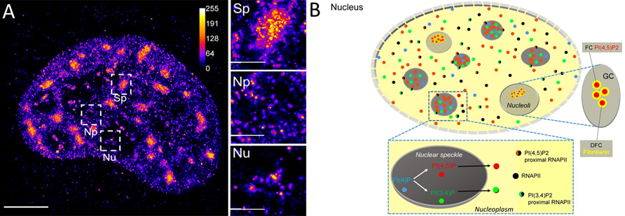
Functional nuclear sub-compartments linked with the crucial cellular processes, such as gene expression, contain phosphoinositides in the form of small foci (Fig. 1). Nevertheless, current models of gene expression largely omit the roles of nuclear lipids and amongst them nuclear phosphatidylinositol phosphates. We aim to fill this gap, and to this end, we use a combination of advanced light and electron microscopy (Fig. 2) together with biochemistry and molecular biology. These approaches allow us to gain insight into the role of nuclear phosphatidylinositol phosphates in the regulation of gene expression and push the boundaries of the field. We identified a novel type of nuclear structures – nuclear lipid islets – that contain phospholipids, proteins, and nucleic acids and serve as platforms for efficient transcription (Sobol et al., 2018). Our efforts continue to systematically unravel the phospholipid identity of the gene expression compartments (Hoboth et al., 2021), and we are testing the idea that PIs and their interactors regulate, at the molecular level, subsequent stages of RNAPII transcription (Sztacho et al., 2021). Moreover, our group has identified nuclear myosin I as a novel transcription factor (Philimonenko et al., 2004) and also contributed to understanding the role of nuclear actin and actin-related proteins in the establishment of functional nuclear architecture and regulation of gene expression (Balaban et al., 2021). Our research centred on the role of nuclear lipids in the formation of lipo-ribonucleoprotein transcription hubs is important for better understanding the role of functional nuclear architecture, nucleoskeleton, and nuclear lipids in the gene expression in health and disease.