Institute of Molecular Genetics of the Czech Academy of Sciences
In the Laboratory of Cell Motility, we study the eukaryotic flagellum and cilium (the terms are interchangeable), a fascinating organelle with motile, signalling and sensory roles. The flagellum/cilium is evolutionarily well conserved and is present on surfaces of many eukaryotic cells, including important parasitic protists and most mammalian cell types. In humans, malfunction of cilia causes severe hereditary disorders called ciliopathies.
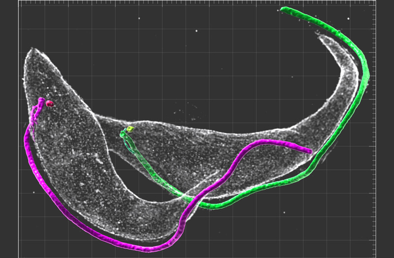
The principal structural and force-generating component of the flagellum/cilium is the microtubule-based axoneme. The axoneme consists of several hundred protein species organized in a highly defined manner. How does the cell form such a complex yet highly organized structure?
Using the highly experimentally tractable flagellated protist Trypanosoma brucei, the causative agent of human African trypanosomiasis, we have developed and optimized biochemical, cell biology, and imaging techniques to identify proteins critical for the processes of axonemal construction and length regulation.
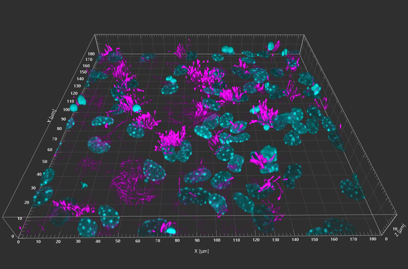
Importantly, due to the high evolutionary conservation of the organelle, we were able to identify mammalian orthologues of some of these proteins and assess their roles in mammalian cells.
Finally, to reveal intrinsic activities of these newly identified proteins, we study their behaviour by in vitro biochemical assays. In particular, we employ microscopy-based single-molecule assays, which provide deep mechanistic insights into the activities of individual molecules.
We believe that integrating these approaches will provide a comprehensive understanding of the processes orchestrating the axonemal growth and will give insights into the causes of certain ciliopathies.