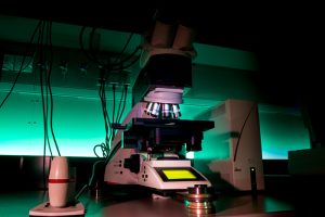
Upright routine widefield fluorescence microscope with motorized stage, fully automated transmitted and fluorescence light axis, and both monochromatic and color cameras. The monochromatic one is a high resolution very sensitive grayscale sCMOS camera. The LAS X Navigator tool allows to create high-resolution image tile-scans of large areas using the precise motorized stage.
| Methods | Routine fluorescence imaging True color RGB imaging in transmitted light mode Brightfield/Darkfield and Phase contrast Large tile-scans & auto merge in LAS X Navigator |
| Illumination | HLX 100 W Halogen lamp for transmitted light HXP 120W/45C VIS Hg lamp – Leica EL6000 for fluorescence |
| Objectives | HC PL FLUOTAR 5x/0.15 DRY; FWD 12 | BF, DF, POL HC PL APO 10x/0.40 DRY PH1; FWD 2.2; CG 0.17 | BF, DF, POL, PH HC PLAN APO 20x/0.70 DRY PH2; FWD 0.59; CG 0.17 | BF, DF, POL, PH HCX PL APO 40x/0.75 DRY PH2; CG 0.17 | BF, DF, POL, PH HCX PL APO 63x/1.40 OIL PH3 CS; FWD 0.1; CG 0.17 | BF, POL, PH HCX PL APO 100x/1.40-0.70 OIL; FWD 0.09; CG 0.17 | BF, DF, POL |
| Filtercubes | DAPI (A4) (Ex: 360/40; DM 400; Em: 470/40) FITC (L5) (Ex: 480/40; DM 505; Em: 527/30) TRITC (N3) (Ex: 546/12; DM 565; Em: 600/40) Cy5 (Y5)(Ex: 620/60; DM 660; Em: 700/75) |
| Cameras | Leica DFC490 – color CCD camera; 2,7 µm pixel Leica DFC 9000 – monochromatic sCMOS camera; 6,5 µm pixel, QE: min. 82 % |
| Stage | motorized microscope stage xyz |
| Software | LAS X, 64 Bit |
| Location | room no. 0.172 (Green) |
| Phone | ext. 3172 |
| Booking | Calpendo (“DM6000” ) |
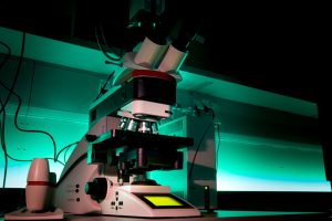
Upright widefield fluorescence microscope with motorized stage, fully automated transmitted and fluorescence light axis and high-resolution very sensitive monochromatic 16-bit sCMOS camera. The LAS X Navigator tool allows to create high-resolution image tile-scans of large areas using the precise motorized stage.
| Methods | Routine fluorescence imaging Brightfield/Darkfield and Phase contrast Large tile-scans & auto merge in LAS X Navigator |
| Illumination | HLX 100 W Halogen lamp for transmitted light HXP 120W/45C VIS Hg lamp – Leica EL6000 for fluorescence |
| Objectives | HC PL FLUOTAR 6.3x/0.13 DRY; FWD 12.860 | BF, POL HC PL FLUOTAR 10x/0.30 DRY PH1; FWD 11.0; CG 0.17 | BF, DF, POL, PH HC PL FLUOTAR 20x/0.40 DRY PH1; FWD 7.5; CG 0.17 | BF, DF, POL, PH HC PLAN APO 20x/0.70 DRY PH2; FWD 0.59; CG 0.17 | BF, DF, POL, PH HCX PL APO 40x/0.75 DRY PH2; FWD 0.28 CG 0.17 | BF, DF, POL, PH HCX PL APO 63x/1.40 OIL; FWD 0.14; CG 0.17 | BF, POL, PH HCX PL FLUOTAR 100x/1.3 OIL PH3; FWD 0.13; CG 0.17 | BF, POL |
| Filtercubes | DAPI (A) (Ex: 360/40; DM 400; Em: LP 425) GFP (Ex: 470/40; DM 495; Em: 525/50) TRITC (N3) (Ex: 546/12; DM 565; Em: 600/40) Cy5 (Y5)(Ex: 620/60; DM 660; Em: 700/75) |
| Cameras | Leica DFC 9000 – monochromatic sCMOS camera; 6,5 µm pixel, QE: min. 82 % |
| Stage | motorized microscope stage xyz |
| Software | LAS X, 64 Bit |
| Location | room no. 0.172 (Green) |
| Phone | ext. 3172 |
| Booking | Calpendo (“DM6000-2”) |
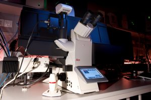
Leica DM8 – Inverted widefield fluorescence microscope with motorized stage and fully automated transmitted and fluorescence light axis. The system is dedicated for live cell imaging. Long-term time-lapse live cell experiments are secured with hardware autofocus system. The microcope is also equipped with infinity port laser-scanner for precise photo-kinetic experiment (FRAP, iFRAP, Photo-activation…). All the features are fully implemented in LAS X software.
For long-term live cell experiments are secured with hardware autofocus system.
| Methods | Routine fluorescence imaging True color RGB imaging in transmitted light mode Brightfield/Darkfield, Phase contrast, Modulation contrast Large tile-scans & auto merge in LAS X Navigator Time-lapse experiments with HW autofocus Multi-positions experiment Photo-kinetic experiments |
| Illumination | LED light source for transmitted light lasers for photomanipulation:
|
| Objectives | HC PL FLUOTAR 5x/0.25, FWD 12 | BF, POL, PH, IMC N PLAN 10x/0.25 DRY; FWD 17.6 | BF, POL, PH, IMC HCX PL FLUOTAR L 20x/0.40 DRY CORR; FWD 6.9; CG 0-2 | BF, POL, PH, IMC HCX PL FLUOTAR 40x/0.95 DRY CORR; FWD 3.3; CG 0-2 | BF, POL, PH, IMC HCX PL APO 40x/1.25 oil | BF, POL HCX PL APO 63x/1.40-0.60 OIL; FWD 0.1; CG 0.17 | BF, POLBF, POL |
| Filtercubes | DAPI (A4) (Ex: 350/50; DM 400; Em: 460/50) FITC (Ex: 480/40; DM 505; Em: 527/30) TRITC (Ex: 546/10; DM 560; Em: 585/40) Y5 (Cy5) (Ex: 620/60; DM 660; Em: 700/75) |
| Cameras | Leica DFC490 – color CCD camera; 2,7 µm pixel Leica DFC 9000 – monochromatic sCMOS camera; 6,5 µm pixel, QE: min. 82 % |
| Stage | motorized microscope stage xyz |
| Aditional equipment | PEACON incubation unit (CO2, temperature, humidity) |
| Software | LAS X |
| Location | room no. 0.171 (Orange) |
| Phone | ext. 3171 |
| Booking | Calpendo (“DMI 8” ) |
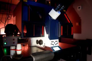
Inverted widefield fluorescence microscope DMI6000 with fully automated transmitted and fluorescence light axis. The transmitted light equipment includes differential interference contrast (DIC) prism. An incubation chamber makes the system suitable for live-cell imaging. The total internal reflection fluorescence illumination module (Leica AM TIRF MC) controls the angle of the excitation beam and makes use of the total reflection to excite only molecules in the thin section in contact with the coverglass. The system is equipped with both FRAP and FRET modules.
| Methods | Routine fluorescence imaging Brightfield, Nomarski contrast (DIC) Large tile-scans & auto merge in LAS X Navigator Multi-positions experiment Time-lapse experiments TIRF microscopy experiments |
| Illumination | HLX 100 W Halogen lamp for transmitted light HXP 120W/45C VIS Hg lamp – Leica EL6000 with for fluorescence solid-state TIRF laser 405 nm solid-state TIRF laser 488 nm solid-state TIRF laser 561 nm solid-state TIRF laser 633 nm |
| Objectives | HCX PL FLUOTAR 10x/0.30 DRY PH1; FWD 11.0 | BF, POL HCX PL FLUOTAR L 20x/0.40 CORR DRY PH1; FWD 6.9; CG 0-2 | BF, POL N PLAN L 40x/0.55 CORR DRY; FWD 3.3; CG 0-2 | BF, POL HCX PL APO 63x/1.3 GLYC CORR 37°C; FWD 0.28; CG 0.14-0.18 | BF, POL, DIC HCX PL APO 63x/1.40-0,60 OIL CS; FWD 0.1; CG 0.17 | BF, POL, DIC HCX PL APO 100x/1.46 OIL CORR CS; FWD 0.09; CG 0.1-0.22 | BF, POL, DIC |
| Filtercubes | A (Ex: 360/40; DM 400; Em: LP425) CFP-T (Ex: 422/44; DM 455; Em: 480/40) GFP-T (Ex: 490/20; DM500; Em: 525/50) YFP-T (Ex: 490/20, Em: 535/30) Cy3-T (Ex: 560/10, Em: 610/65) Cy5-T (Ex: 635/10, Em: 720/60) TRI (B/G/R) (ExFW: 420/30; 495/15; 570/20, Em: 457/22;530/20;628/28) FRET kostka pro CFP/YFP (ExFW: 436/12, 500/20; EmFW: 467/37, 545/45) QUAD-T (ExFW: 405/12, 490/20, 560/30,635/20; EmFW: 450/50, 525/36,600/32,705/72) |
| Cameras | Leica DFC350FX R2 – monochromatic CCD camera; 6,4 µm pixel Leica DF300 FX – color CCD camera; 6,4 µm pixel Andor iXon 897 – high sensitivity EMCCD back-iluminated camera, 512×512 px, suitable for single molecule live cell imaging; 16 µm pixel |
| Stage | motorized microscope stage with Super Z-galvo scanning insert for fast and precise Z movement |
| Aditional equipment | incubation unit (CO2, temperature, humidity) acousto-optical tunable filter (AOTF) – laser control |
| Software | LAS X – 64 bit; with FRET and FRAP and deconvolution modules |
| Location | room no. 0.171 (Orange) |
| Phone | ext. 3171 |
| Booking | Calpendo (“TIRF”) |
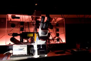
The system is built on the inverted widefield Olympus microscope version IX-71. The system is equipped with the HW autofocus system and an incubation chamber, and is suitable for multi-location and time-lapse experiments. The lasers for photomanipulation enable performance of photokinetics experiments (FRAP) and FRET. The sensitive camera makes the microscope suitable for live cell imaging and low fluorescence intensity applications.
| Methods | Routine fluorescence imaging Brightfield, Nomarski contrast (DIC) Tile-scans Multi-positions experiment Time-lapse experiments with HW autofocus Photo-kinetic experiments |
| Illumination | Lumencore LED – solid-state light source for fluorescence lamp for transmitted light lasers for photomanipulation (adjustable spot size):
|
| Objectives | U PLAN FL 20x/0.50 DRY PH1; FWD 1.6; CG 0.17 | BF, POL U PLAN FL 40x/0.75 DRY; FWD 0.51; CG 0.17 | BF, POL U APO/340 40x/1.35-0.65 CORR OIL; FWD 0.1; CG 0.17 | BF, POL PLAN APO N 60x/1.42 OIL; FWD 0.15; CG 0.17 | BF, POL, DIC |
| Lumencore excitation filters | DAPI 390/18 CFP 438/24 GFP/FITC 475/28 YFP 513/17 RD-TR-PE 542/27 mCherry/Alexa594 575/25 Cy5 632/22 |
| Dichroic mirrors | Standard (DAPI, FITC, RD-TR-PE, Cy5) Live Cell (CFP, YFP, mCherry) Alexa (GFP, mCherry, Alexa 594) |
| Emission filter wheel | DAPI (435/48) FITC (523/36) RD-TR-PE (594/45) Alexa 594 (632/60) Cy5 (676/34) GFP (525/50) mCherry (632/60) CFP (470/24) YFP (559/38) |
| Eyepiece filers | the same as on emission filter wheel (excluding Cy5 filter) |
| Cameras | Photometrics CoolSANP HQ – high sensitive CCD camera, 1024×1024 px; 6,45 µm pixel |
| Stage | motorized stage with xyz movement equipped with HW AF |
| Aditional equipment | incubation unit (CO2, temperature, humidity) antivibration table hardware Ultimate autofocus for maintenance in-focus stability within the entire specimen |
| Software | SoftWorx
|
| Location | room no. 0.171 (Orange) |
| Phone | ext. 3171 |
| Booking | Calpendo (“DVcore”) |
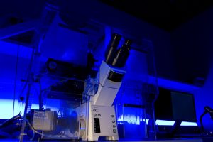
Scan^R is a module built on the inverted widefield microscope Olympus IX81. The system is designed for fully automated image acquisition and data analysis of biological samples. Scan^R can handle many different specimen formats, including multi-well plates, slides and custom-built arrays. The hardware autofocus maintains in-focus stability within the entire specimen. The acquisition is followed by complex image analysis and advanced data evaluation. Due to an incubation unit and a sensitive camera, the microscope is suitable for live cell imaging and low fluorescence intensity applications.
| Methods | High-content imaging (preferentially in widefield fluorescence mode) |
| Illumination | Xenon Mercury mixed gas arc lamp MT20 150 W (intense peaks at 365, 405, 436, 546, 577) lamp for transmitted light |
| Objectives | LUCPLFN 20x/0.45 DRY CORR PH1; FWD 6.6-7.8; CG 0-2 LUCPLFN 40x/0.6 DRY CORR PH2; FWD 2.7-4; CG 0-2 UPLSAPO 10x/0.4 DRY; FWD 3.1; CG 0.17 UPLFLN 40x/1.3 OIL; FWD 0.2; CG 0.17 UPLSAPO 40x/0.95 DRY CORR; FWD 0.18; CG 0.11 – 0.2 UPLSAPO 60x/1.35 OIL; FWD 0.15; 0.17 Optional Tube lens 1.6x |
| Excitation filters | DAPI 360/40 nm FITC 487/15 nm TRITC 552/25 nm GFP 470/20 nm HQ Cy5 623/55 nm CFP 435/10 nm YFP 500/20 nm |
| Emission filtercubes | DAPI/FITC/TRITC (460/10; 522/15; 605/30 nm) GFP HQ (518/45 nm HQ) Cy5 (705/35 nm) CFP/YFP (470/20; 545/50 nm) |
| Cameras | sCMOS camera Hammamatsu ORCA-Flash4.0 V2 – 6,5 µm pixel |
| Stage | motorized microscope stage xyz |
| Aditional equipment | incubation unit (CO2, temperature, humidity) antivibration table hardware autofocus |
| Software | ScanR acquisition ScanR analysis |
| Location | room no. 0.175 (Blue) |
| Phone | ext. 2750 |
| Booking | Calpendo (“ScanR”) |
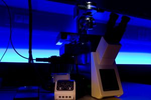
Scan^R is a module built on the inverted widefield microscope Olympus IX81. The system is designed for fully automated image acquisition and data analysis of biological samples. Scan^R can handle many different specimen formats, including multi-well plates, slides and custom-built arrays. The hardware autofocus maintains in-focus stability within the entire specimen. The acquisition is followed by complex image analysis and advanced data evaluation. Due to an incubation unit and a sensitive camera, the microscope is suitable for live cell imaging and low fluorescence intensity applications.
| Methods | High-content imaging (preferentially in widefield fluorescence mode) |
| Illumination | Lumencore LED – solid-state light source for fluorescence (peaks: 395, 438, 475, 511, 555, 575 a 635 nm) Lamp for transmitted light |
| Objectives | UPLSAPO 10x/0.4 DRY; FWD 3.1; CG 0.17 UPLXAPO 20x/0.8 DRY CORR; FWD 0.6; CG 0 – 2 UPLXAPO 40x/0.95 DRY CORR; FWD 0.18; CG 0.11 – 0.2 UPLXAPO 60x/1.42 OIL; FWD 0.15; 0.15 Plan apochromatic 20x, NA 0.8, WD 0,6 mm |
| Excitation LED source | DAPI 395/25 nm, CFP 438/29 nm, FITC 475/28 nm, YFP 511/16 nm, TRITC 555/28 nm mCherry 575/25 nm Cy5 635/22 nm |
| Emission filtercubes | QUAD dichroic: DAPI/FITC/Cy3/Cy5 |
| Cameras | sCMOS camera Hammamatsu ORCA-Flash4.0 LT+90, pixel 6,5 µm pixel |
| Stage | motorized microscope stage xyz |
| Aditional equipment | hardware autofocus |
| Software | ScanR acquisition ScanR analysis |
| Location | room no. 0.175 (Blue) |
| Phone | ext. 2750 |
| Booking | Calpendo (“ScanR-2”) |
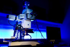
Zeiss Axiom Zoom.V16 – Fully motorized fluorescence macroscope with high resolution objectives is equipped with two cameras and a motorized 16:1 zoom with apochromatic correction. The system is well suited for a wide range of bigger samples and has superior brightness in large object fields. ApoTome.2 uses structured illumination to improve contrast and to increase resolution of the optical sections.
| Methods | Fluorescence imaging on widefield OR Apotome (optical sectioning) mode Transmitted and/or reflected light True color RGB imaging in transmitted light mode Brightfield/Darkfield, Opaque contrast Large tile-scans & auto merge in ZEN Blue software Time-lapse experiments Multi-positions experiment |
| Illumination | HXP 120 V – metal halid lamp for fluorescence CL 9000 LED CAN – transmitted light source CL 9000 LED CAN – incident light source |
| Objectives | PlanNeoFluar Z 1.0x/0.25; FWD 56 | BF, DF, oblique illumination + Reflected light PlanNeoFluar Z 2.3x/0.57; FWD 10.6 | BF, DF, oblique illumination + Reflected light |
| Fluorescence filtercubes | DAPI Filter set 49 (Ex: G 365; BS FT 395; Em: BP 445/50) eGFP Filter set 38 HE (Ex: BP 470/40; BS FT 495; Em: BP 525/50) Cy3 Filter set 63 HE (Ex: BP 572/25; BS FT 590; Em: BP 629/62) Cy5 Filter set 50 (Ex: BP 640/30; BS FT 660; Em: BP 690/50) |
| Cameras | ZEISS Axiocam 305 – color CMOS camera; 3,45 µm pixel ZEISS Axiocam 512 mono – monochromatic CCD camera; 3,1 µm pixel |
| Additional equipment | ApoTome.2 for optical sectioning |
| Software | ZEN Blue 2.3 |
| Location | room no. 0.175 (Blue) |
| Phone | ext. 2750 |
| Booking | Calpendo (“Apotome”) |
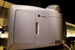
Z.1 light sheet fluorescence microscope allows fast, gentle multi-view imaging of large specimens. The microscope enables to record the development of living specimens and to deliver exceptionally high information content which includes the inner structures. To image deep into fixed non-transparent biological samples, it is necessary to apply a tissue clearing protocol (e.g. Scale, FocusClear, CUBIC).
| Methods | Fluorescence imaging with lightsheet optical sectioning Dual-site & multi-field scanning, processing at ZEN Black software Time-lapse experiments Multi-positions experiment |
| Illumination | 405nm Solid-state laser, 50mW 445nm Solid-state laser, 25mW 488nm Solid-state laser, 30mW 561nm Solid-state laser, 20mW 638nm Solid-state laser, 75mW Infrared LED light source for trasmitted light |
| Ilumination objectives | Objective Lightsheet Z.1 5x/0.1 Objective Lightsheet Z.1 10x/0.2 |
| Detection objectives | Dry objective Lightsheet Z.1 5x/0.16 Immersion objective Lightsheet Z.1 10x/0.5 Immersion objective Lightsheet Z.1 20x/1.0 Immersion objective Lightsheet Z.1 40x/1.0 Objective Clr Plan-Apochromat 20x/1.0 Corr nd=1,38 (suited for clearing media with RI 1.38, e.g. Scale) Objective Clr Plan-Neofluar 20x/1.0 Corr nd=1.45 (suited for clearing media with RI 1.45, e.g. FocusClear) |
| Laser blocking filters | 405/488/561 tripple excitation dichroic 405/488/561/640 quad excitation dichroic 405/488/640 tripple excitation dichroic 445/561 dual excitation dichroic |
| Emission filter sets | DAPI-GFP (SBS LP 490; Em: BP 420-470 + BP 505-545) DAPI-Cy3 (SBS LP 510; Em: BP 420-470 + BP 575-615) GFP-Cy3 (SBS LP 560; Em: BP 505-545 + BP 575-615) GFP-mCherry (SBS LP 560; Em: BP 505-545 + LP 585) GFP-DRAQ5 (SBS LP 560; Em: BP 505-545 + LP 660) GFP narrow-mCherry (SBS LP 560; Em: BP 505-530 + LP 585) |
| Cameras | camera PCO.Edge 5.5 (sCMOS) – 2 pieces; 6,5 µm pixel |
| Additional equipment | incubation unit (CO2, temperature, humidity) antivibration table for SPIM microscopy PC for storage and data analysis stereomicroscope for sample preparation and system maintenance (Stemi 305 EDU Microscope Set) |
| Software | ZEN Black system 2014 for Lightsheet Z.1 |
| Location | room no. 0.173 (Yellow) |
| Phone | ext. 2750 |
| Booking | Calpendo (“Z1”) |
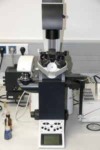
Leica DMI4000 B – Inverted widefield fluorescence microscope with manual ZX stage, manual objective Z drive and manual objective turret. This microscope is for routine simple applications only. NOT RECOMMENDED FOR IMAGE ACQUISITION. ACQUISITION FEATURES ARE NOT FULLY SUPPORTED.
| Methods | Fluorescence and transmitted light observation (imaging is NOT supported) |
| Illumination | Leica Xenon lamp (75 W) for fluorescence lamp for transmitted light |
| Objectives | N PLAN 10x/0.25 DRY; FWD 17.6 N PLAN L 40x/0.55 CORR DRY; FWD 3.3; CG 0-2 PL APO 63x/1.40 oil |
| Fluorescence filtercubes | A (Ex: 360/40; DM 400; Em: LP425) CFP (Ex: 436/20; DM 455; Em: 480/40) GFP (Ex: 470/40; DM 500; Em: 525/50) YFP (Ex: 500/20; DM 520; Em: 535/30) RFP (Ex: 546/12; DM 560; Em: 605/75) Cy5 (Y5) (Ex: 620/60; DM 660; Em: 700/75) |
| Cameras | Leica DC350FX – monochromatic CCD camera; 6,4 µm pixel |
| Software | LAS X |
| Location | room no. 2.38 |
| Phone | ext. 3106 |
| Booking | reservation not needed |
