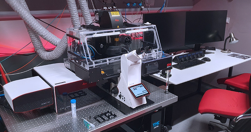
Fully motorized inverted microscope DMi8 with confocal head Leica STELLARIS 8 equipped with wide-range white light laser with pulse picker (WLL PP), highly sensitive time-resolved detection, FCS, and STED is a top-of-the-line system for a variety of high-end applications. The system is equipped with several acquisition modules for photo-kinetic experiments, FLIM or FLIM-FRET based experiments, the high-resolution imaging followed with Lightning adaptive deconvolution module, and FCS and FCCS modules for studying molecular dynamics. Furthermore, it contains a STED module with a TauSTED Xtend which can reach normal STED resolution with much lower depletion laser power, making it uniquely suited for super-resolution acquisitions of living cells. The system supports AI-based workflow for fast rare event detection using Autonomous Microscopy powered by Aivia software.
The combination of white light laser, AOBS and spectral sliders on detector cascade allows the fine tuning of excitation laser line selection and precise definition of emission spectra.
Stability of long-term live cell imaging experiments is ensured by a hardware autofocus system (AFC) and OKOLAB incubation system, which provides temperature, CO2, and humidity control.
| Methods | TauSTED Xtend – gentle fluorescence lifetime-based STED Confocal scanning fluorescence imaging with Lightning super-resolution module Transmitted light imaging Brightfield and Nomarski contrast (DIC) (automated) Tile-scans & merge in LAS X software Time-lapse experiments Multi-positions experiments Photo-kinetic experiments FRET (SE or Acceptor photobleaching) FLIM, FRET-FLIM experiments Rare event detection and Autonomous microscopy using Aivia software |
| Illumination | Excitation lasers: 405 nm CW diode laser, AOTF modulated, 100mW 440 – 790 nm Pulsed white light laser with pulse picker (WLL PP): – Range of wavelengths selection: 440 – 790 nm – Laser line selection step: 1 nm – Laser power:> 0,9 mW 440-485 nm,> 1,8 mW 485-790 nm – Possible laser frequencies: 80, 40, 20, 10, 5, 2,5 MHz – 8 channel AOTF – WLL PP in combination with AOBS allows up to 8 independent parallel laser lines Depletion lasers: – 589 nm pulsed STED, >1.5 W – 775 nm pulsed STED, >1.5 W 12V 100W LED source for transmitted light HXP 120W/45C VIS Hg lamp – Leica EL6000 for fluorescence |
| Objectives | HC PL FLUOTAR 10x/0.30 DRY CS2; FWD 11.0 mm | BF, POL, DIC HC PL APO 20x/0.75 IMM CORR CS2; FWD 0.67 mm | BF, POL HC PL APO 93x/1,30 GLYC motCORR STED WHITE; FWD 0.30 mm| BF, POL, DIC PL APO 40x/1.30 OIL CS2; FWD 0.24; CG 0.17 | BF, POL, DIC HC PL APO 63x/1.40 OIL CS2; FWD 0.14 MM| BF, POL, DIC HC HC PL APO 100x/1.4 OIL STED WHITE CS2; FWD 0.13; CG 0.17 | BF, POL, DIC (OPTIONAL – UPON REQUEST) |
| Eyepiece filtercubes | DAPI (Ex: 355/56, DC: 400, Em: 460/50) GFP (Ex: 470/40; DC 495; Em: 525/50) RHOD (Ex: 546/10; DC 560; Em: 585/40) CFP/YFP (Ex: 435/25 + 500/20; DC 450 + 515; Em: 470/25 + 535/30) |
| Detectors | 2x supersensitive silicon-based hybrid detectors (HyD-S) 2x supersensitive hybrid detectors (HyD-X) for very fast time-resolve acquisition (FLIM) 1x supersensitive hybrid detector (HyD-R) for long wavelengths (700 – 850 nm) 1x transmitted light TL – PMT detector |
| Confocal head | 8-channels acousto-optical tunable filter (AOTF) for fast laser intensity modulation Acousto-optical beam splitter (AOBS) Optical rotation Conventional linear scanner (1-2600 Hz line frequency) Maximum scanner resolution 8192×8192 pixels Hardware zoom 0.75x-48x Scanned field size 22 mm (diagonally) Resonant scanner (8000 Hz line frequency) Maximum scanner resolution 2496 x 2496 pixels Hardware zoom 1.25x-48x |
| Time-resolve modules | Tau-sense modules for time-resolved acquisition (time-resolved imaging without further analysis): – TauGating – TauContrast – TauSeparation STELLARIS FALCON: FAST Lifetime CONtrast (FALCON) is solution for fast image acquisition, processing and FLIM analysis with modules: – FLIM component fitting – FLIM Phasor diagram – FLIM-FRET analysis |
| Computational super-resolution module | Lightning – a super-resolution module based on mutual acquisition control and adaptive deconvolution based on SNR decision mask estimation. Maximal declared resolution is 120 nm laterally and 200 nm axially. |
| Stage | Motorized microscope stage with Super Z-galvo scanning insert for fast and precise Z movement |
| Additional equipment | Antivibration table OKO-LAB incubation unit (CO2 + O2, temperature and humidity control system) |
| Software | All features are fully implemented to the LAS X software LAS X modules: LAS X LiveDataMode LAS X 3D Visualisation LAS X Phasor LAS X FRAP, FRAP Zoomer LAS X Assay (Navigator) LAS X Lightning LAS X Navigator Expert LAS X Rare Event Detection Aivia |
| Location | room no. 0.174 (Red) |
| Phone | ext. 2433 |
| Booking | Calpendo (“Stellaris”) |

The DeltaVision OMX imaging platform is an advanced multi-mode, super-resolution inverted microscope system which offers super-resolution imaging using 3D Structured Illumination (3D-SIM) and Dense Stochastic Sampling Imaging (DSSI) – Localization Microscopy. The Blaze SIM Module offers dynamic high speed 3D-SIM suitable for live cell super-resolution imaging. Blaze incorporates a proprietary, ultra-fast, structured illumination module, advanced optical platform design and the latest high-speed camera technologies. The system can be used also for super-fast widefield acquisition and photo-kinetic experiments (FRAP).
Methods
Illumination
lamp for transmitted light
InsightSSI (Lumencore) illumination module – for WF imaging; standard shutters, open/close time ~ 1.8 ms excitation lasers – for 3D SIM, TIRF and localization; high speed galvanometer controlled shutters, open/close time ~ 200 μs
3D-SIM resolution at different wavelengths
| Laser | Type | Power (mW) | Expected XY resolution | Expected Z resolution |
| 405 nm | diode | 100 ± 10 | 110 ± 5 nm | 340 ± 10 nm |
| 445 nm | diode | 100 ± 5 | 115 ± 5 nm | 340 ± 10 nm |
| 488 nm | OPSL | 100 ± 4 | 120 ± 5 nm | 340 ± 10 nm |
| 514 nm | OPSL | 100 ± 6 | 120 ± 5 nm | 350 ± 10 nm |
| 568 nm | OPSL | 100 ± 4 | 135 ± 5 nm | 350 ± 10 nm |
| 642 nm | diode | 110 ± 10 | 160 ± 5 nm | 380 ± 20 nm |
Objectives
PLAN APO N 60x/1.42 OIL; FWD 0.15; CG 0.17 | BF, POL, DIC (optimized)
U APO N 60x/1.49 CORR OIL TIRF; FWD 0.1; CG 0.13-0.19 & 23/37°C | BF, POL, DIC
U APO N 100x/1.49 CORR OIL TIRF; FWD 0.1; CG 0.13-0.19 & 23/37°C | BF, POL, DIC
U PLAN S APO 60x/1.3 CORR SILICONE; FWD 0.3; CG 0.15-0.19 & 23/37°C | BF, POL, DIC
InsightSSI excitation filters (for widefield applications only)
DAPI 395.5/29
FITC 477/32
mCherry/Alexa Fluor 568 571/19
Cy5 645.5/15
CFP 438/24
YFP 512.5/15
Emission filters
DAPI 435.5/31
FITC 528/48
mCherry/Alexa Fluor 568 609/37
Cy5 683/40
CFP 477.5/35
YFP 541/22
Cameras
4x pco.edge 5.5 sCMOS, Readout speeds 95 MHz, 286 Mhz, 15 bit, pixel size: 6.5 μm camera, 80 nm 60x, 48 nm 100x
Stage
XYZ nanomover and fast Z-piezo
Additional equipment
antivibration table
sample holders:
Software
OMX Acquisition control software running on OMX Master computer under Windows 7
SoftWoRx – image reconstruction, deconvolution and analysis running on the SI workstation running under the CentOS v6 (Linux)
Dense Stochastic Sampling Imaging (DSSI) algorithm for localization microscopy images
Location: room no. 0.174 (Red)
Phone: ext. 2433
Booking: Calpendo (“OMX”)

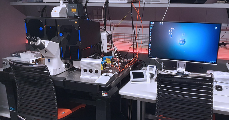
The Elyra 7 is a motorized inverted microscope equipped with a super-resolution system that uses a specialized lattice illumination pattern for 3D structured illumination (SIM) images and single-molecule localization methods (SMLM) like STORM and PALM. The lattice illumination allows for faster, gentler, and deeper imaging of live samples, and can reach up to 255 FPS during time-lapse acquisition. The unit has an incubation chamber for live imaging with controlled CO2 and temperature. The signal detection is achieved through two sCMOS cameras which enable simultaneous detection of two different fluorophores.
| Methods | Lattice SIM, SIM2 Single Molecule Localization Microscopy (3D PALM / dSTORM) TIRF Apotome (optical sectioning) mode Transmitted light imaging Brightfield and Nomarski contrast (DIC) (automatic) Time-lapse experiments (live-cell imaging) Multi-positions experiments |
| Illumination | Halogen 12 V/100W lamp – for transmitted light Metal halide HXP 120 V lamp – for fluorescence Excitation lasers (for TIRF, SIM, SMLM): – 405 nm diode, 50 mW – 488 nm OPSL, 500 mW – 561 nm OPSL, 500 mW – 642 nm OPSL, 500 mW |
| Objectives | EC Plan-Neofluar 10x/0.3 M27 DRY | BF, POL C-Apochromat 40x/1.2 Water Corr M27 | BF C-Apochromat 63x/1.2 Water Corr M27 | BF, DIC α Plan-Apochromat 63x/1.46 Oil Corr M27 | BF, TIRF α Plan-Apochromat 100x/1,46 Oil DIC M27 | BF, DIC, TIRF |
| Eyepiece filtercubes | Laser Blocking Filter for 405/488/561/642 named: – DAPI – FITC – mCherry – Cy5 Simultaneous two-channel viewing: – GFP + mCherry – GFP + Cy5 Sequential four-channel viewing: – DAPI and mCherry + GFP and Cy5 – DAPI and GFP + mCherry and Cy5 |
| Cameras | DuoLink for a parallel imaging on two cameras: – 2x pco.edge sCMOS (version 4.2 CL HS) – thermal stabilization at 0°C / +5 °C / +7 °C – 16-bit depth – high resolution: 2048 x 2048 px – pixel size 6.5 x 6.5 µm – quantum efficiency up to 82% – exposure times from 100 µs to 20 s – maximum frame rate 100 fps |
| Superresolution modules | Lattice SIM, SIM2 Single Molecule Localization Microscopy (3D PALM / dSTORM) |
| Stage | XY Scanning Stage Piezo 130 x 100 (D): – Movement range 130 mm x 100 mm (adjustable) – Max speed 100 mm/s – 0.2 μm resolution – Repeatability +/- 1 μm – Absolute accuracy +/- 5 μm Z-piezo insert: – Movement range at least 100 μm – < 5 nm resolution – Suitable for: Standard microscopic slip: 76 x 26 mm, Petri dish 30 – 35 mm in diameter, LabTek II chamber, 55 x 24 mm |
| Additional equipment | Antivibration table 120 x 90 cm Definite Focus – holding focus to compensate Z drift Dark incubation unit (CO2, temperature < 40°C, humidity) – Imaging chamber for Z-piezo inserts PC for storage and data analysis |
| Software | ZEN 3.0 (black edition) 64-bit Modules: – Lattice SIM2/SIM2 Apotome – SMLM (PALM/dSTORM) module – 3D-PALM – Measurement – Multi Channel – Panorama – Manual Extended Focus – Image Analysis – Time Lapse – Tiles & Positions – Z-stack – Extended Focus – Autofocus – Colocalization – Connect Entry (ZEN blue edition) |
| Location | room no. 0.171 (Orange) |
| Phone | ext. 3171 |
| Booking | Calpendo (“Elyra 7”) |
