Institute of Molecular Genetics of the Czech Academy of Sciences

Z.1 light sheet fluorescence microscope allows fast, gentle multi-view imaging of large specimens. The microscope enables to record the development of living specimens and to deliver exceptionally high information content which includes the inner structures. To image deep into fixed non-transparent biological samples, it is necessary to apply a tissue clearing protocol (e.g. Scale, FocusClear, CUBIC).
| Methods | Fluorescence imaging with lightsheet optical sectioning Dual-site & multi-field scanning, processing at ZEN Black software Time-lapse experiments Multi-positions experiment |
| Illumination | 405nm Solid-state laser, 50mW 445nm Solid-state laser, 25mW 488nm Solid-state laser, 30mW 561nm Solid-state laser, 20mW 638nm Solid-state laser, 75mW Infrared LED light source for trasmitted light |
| Ilumination objectives | Objective Lightsheet Z.1 5x/0.1 Objective Lightsheet Z.1 10x/0.2 |
| Detection objectives | Dry objective Lightsheet Z.1 2.5x/0.12; FWD 8.7 mm Dry objective Lightsheet Z.1 5x/0.16; FWD 18.5 mm Immersion objective Lightsheet Z.1 10x/0.5; FWD 3.7 mm Immersion objective Lightsheet Z.1 20x/1.0; FWD 5.6 mm Immersion objective Lightsheet Z.1 40x/1.0; FWD 2.5 mm Immersion objective Clr Plan-Apochromat 20x/1.0 Corr nd=1,38; FWD 5.6 mm (suited for clearing media with RI 1.38, e.g. Scale) Immersion objective Clr Plan-Neofluar 20x/1.0 Corr nd=1.45; FWD 5.6 mm (suited for clearing media with RI 1.45, e.g. CUBIC, ECi) |
| Laser blocking filters | 405/488/561 tripple excitation dichroic 405/488/561/640 quad excitation dichroic 405/488/640 tripple excitation dichroic 445/561 dual excitation dichroic |
| Emission filter sets | DAPI-GFP (SBS LP 490; Em: BP 420-470 + BP 505-545) DAPI-Cy3 (SBS LP 510; Em: BP 420-470 + BP 575-615) GFP-Cy3 (SBS LP 560; Em: BP 505-545 + BP 575-615) GFP-mCherry (SBS LP 560; Em: BP 505-545 + LP 585) GFP-DRAQ5 (SBS LP 560; Em: BP 505-545 + LP 660) GFP narrow-mCherry (SBS LP 560; Em: BP 505-530 + LP 585) |
| Cameras | Camera PCO.Edge 5.5 (sCMOS) – 2 pieces; 6,5 µm pixel |
| Additional equipment | Incubation unit (CO2, temperature, humidity) Antivibration table for SPIM microscopy PC for storage and data analysis Stereomicroscope for sample preparation and system maintenance (Stemi 305 EDU Microscope Set) |
| Software | ZEN Black system 2014 for Lightsheet Z.1 |
| Location | room no. 0.173 (Yellow) |
| Phone | ext. 2471 |
| Booking | Calpendo (“Z1”) |
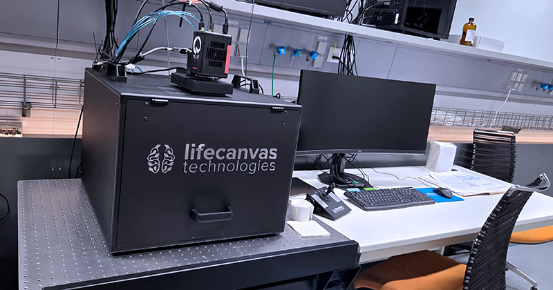
Thanks to uniform axial resolution across the whole sample, rapid volumetric imaging, and a custom imaging chamber, the SmartSPIM light-sheet fluorescent microscope provides the accuracy, speed, and flexibility suitable for imaging of large specimens (e.g. the adult mouse brain or even whole mouse body). The SmartSPIM has been specified to fully automate key microscope features for smooth protocol operation. This includes automation of objective focus control (10nm resolution, 1.75mm/s max speed and 50mm travel range), escape & refocus, optical tuning for immersion refractive index, XYZ sample movement and emission filter selection can all be controlled within SmartSPIM acquisition software. To image deep into fixed non-transparent biological samples, it is necessary to apply a tissue clearing protocol (e.g. iDISCO, SmartBatch+ clearing).
| Methods | Fluorescence imaging with light-sheet optical sectioning Z-stack imaging Dual-site & multi-field scanning (tilescan) Multi-positions experiment |
| Illumination | 488nm Solid-state laser, 150mW 561nm Solid-state laser, 150mW 638nm Solid-state laser, 160mW |
| Ilumination objective | Custom designed, NA=0.125, broadband chromatically corrected (Light-sheet thickness ≤ 8 um with objective 1,6x, 3.8 um with objective 3.6x and ≤ 2,5 µm with objective 9x) |
| Detection objectives | Multi-Immersion (RI=1.33 – 1.56) LCT 1.6x/0.1; FWD 17.5 mm; Pixel size 4 µm; FOV 8 mm Multi-Immersion (RI=1.33 – 1.56) LCT 3.6x/0.2; FWD 17.5 mm; Pixel size 1.8 µm; FOV 3.6 mm Multi-Immersion (RI=1.33 – 1.56) ASI 9x/0.3; FWD 18 mm; Pixel size 0.7 µm; FOV 1.4 mm |
| Laser blocking filters | 488/561/638 tripple excitation dichroic |
| Emission filter sets | GFP – 525/50 mCherry – 600/50 Cy5 – 700/75 |
| Camera | sCMOS camera Hamamatsu ORCA-Fusion BT C15440, 6.5 µm pixel, QE >95%, cooled to -8°C, resolution 2304 x 2304 pixels |
| Additional equipment | Antivibration table for SPIM microscopy PC for acquisition and postprocessing Sample chambers: 60 mm x 118 mm x 60 mm and 80 mm x 174 mm x 60 mm (w x l x h) – sample imaging area up to 40 mm x 70 mm in lateral dimensions |
| Software | SmartSpim acquisition software LifeCanvas Postprocessing: Destripe and Stitching Imaris Viewer |
| Location | room no. 0.173 (Yellow) |
| Phone | ext. 2471 |
| Booking | Calpendo (“SmartSPIM”) |