Institute of Molecular Genetics of the Czech Academy of Sciences
Already next week, the Institute of Molecular Genetics of the Czech Academy of Sciences (IMG) will become the stage for the Czech premiere of the Viventis SCAPE microscope – a brand‑new imaging technology that was introduced to the world only recently and is already reshaping how scientists observe living systems.
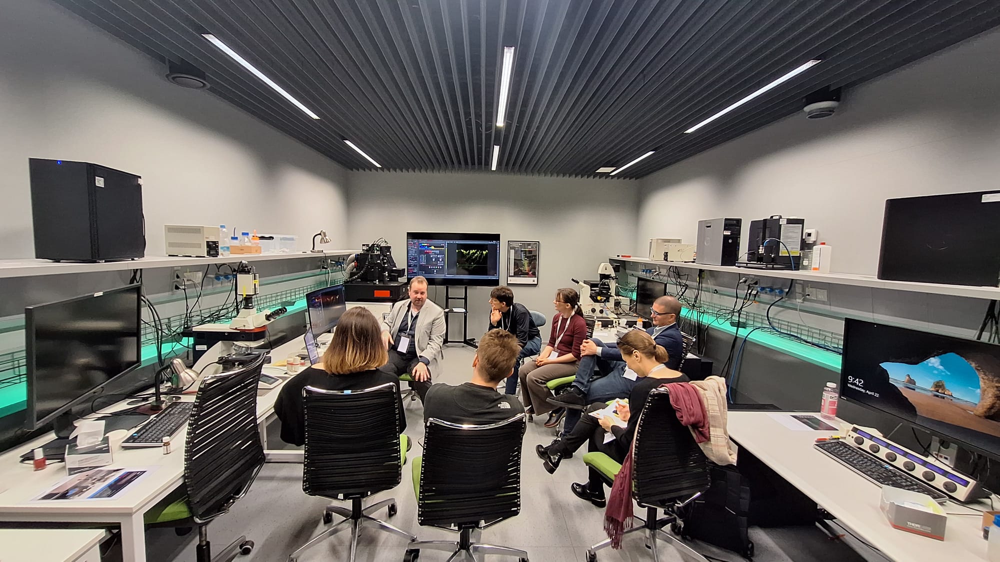
By hosting Vision for Life – Innovation Days in Bioscience Imaging, IMG is bringing this cutting‑edge microscope to the Czech Republic at the earliest possible moment, offering local researchers and microscopy specialists a rare opportunity to explore a technology that has until now been seen mainly at major international conferences.
As IMG Director Petr Dráber emphasizes, the motivation goes far beyond showcasing a new device:
“By hosting the Czech premiere of the Viventis SCAPE microscope at IMG so soon after its global launch, we reaffirm the commitment of our institution and of the large research infrastructure Czech‑BioImaging to continuously support innovation and to provide the most advanced imaging technologies to the Czech and international scientific community.”
The Viventis SCAPE microscope was officially launched at the end of March 2026, during the opening of the Focus on Microscopy (FOM) 2026 conference in Stockholm, Sweden, one of the world’s most important meetings dedicated to advanced microscopy. There, it was introduced as a new generation of light‑sheet microscopy, designed specifically for fast, gentle, three‑dimensional imaging of living samples.
The reaction among scientists working with live cells, tissues, and organoids was immediate. Early users pointed to the system’s ability to capture rapid biological processes without disturbing the natural behavior of cells – a long‑standing challenge in biological imaging.
At first glance, the key principle behind this breakthrough may sound counterintuitive: how can using less light reveal more information? In biology, light is not neutral. Living cells respond to illumination, and excessive light can stress them, alter their behavior, or halt delicate processes altogether. Viventis SCAPE addresses this problem by illuminating only a thin, precisely controlled plane of the sample at any given moment. Everything else remains in the dark. The result is dramatically reduced phototoxicity and photobleaching, allowing cells to behave naturally even during long imaging experiments.
With this technology, microscopy stops being about isolated images and begins to resemble storytelling. Researchers can follow how cells move, stretch, divide, and communicate over time. They can observe tissues reorganize, neurons transmit signals, or tiny beating structures synchronize their rhythms.
As Jiří Černý, an expert in light sheet microscopy at the Light Microscopy Core Facility at IMG, explains:
“With the Viventis SCAPE microscope, the advantages of light‑sheet microscopy become accessible to conventional microscopic specimens. The need for complex mounting of living organisms is eliminated; a completely unique imaging technology makes it possible to capture the entire volume of the sample without moving the specimen—extremely fast and without negative phototoxic effects.”
Instead of reconstructing events after the fact, scientists can now observe cause and effect directly – seeing how one change triggers another and how complex behaviors emerge from countless small interactions. Thanks to compatibility with standard laboratory dishes and multi‑well plates, the system also enables parallel observation of multiple samples, connecting detailed imaging with broader biological questions.
Many of the technologies that reshape science do not emerge overnight. They are born in academic laboratories, refined through years of fundamental research, and gradually mature into tools that transform how we study the living world. The roots of SCAPE microscopy reach back almost 20 years to research at Imperial College London, where physicist Professor Christopher Dunsby developed the principle of single‑objective light‑sheet imaging. For a long time, the method remained confined to highly specialized research labs.
With Viventis SCAPE, Leica Microsystems has translated this academic breakthrough into a robust, user‑friendly system that can be used in everyday research environments – without complex sample mounting or custom‑built setups.
For IMG, presenting this technology is about more than a single event. As Petr Dráber puts it:
“At IMG, we see access to cutting‑edge infrastructure as a long‑term commitment to creating an environment where new scientific questions can be asked and answered. Events like this open the door for researchers, specialists, and young scientists to engage directly with technologies that are shaping the future of life‑science research.”
For researchers, core‑facility specialists, and microscopy enthusiasts, Vision for Life – Innovation Days in Bioscience Imaging offer a rare opportunity to learn directly from experts, explore real data, and imagine experiments that were not previously possible. Sometimes, scientific progress begins not with a new discovery, but with a new way of looking.