Institute of Molecular Genetics of the Czech Academy of Sciences
Supervisor
Martin Gregor
Project description
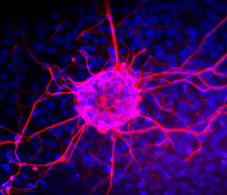
Plectin is a key cytoskeletal crosslinker required for mechanical stability and signaling in skeletal muscle and heart. Mutations in the plectin isoform P1f cause limb-girdle muscular dystrophy accompanied by progressive cardiomyopathy. Due to the large size of full-length plectin, classical gene-replacement strategies are not feasible, necessitating alternative therapeutic approaches.
This PhD project focuses on the development and functional characterization of mini-plectins engineered plectin variants retaining essential functional domains while being compatible with AAV-mediated gene delivery. The main objective is to evaluate their capacity to restore cytoskeletal organization and function in muscle and cardiac cells.
In the in vitro part, the student will analyze expression, localization, and functional integration of miniplectins in cultured cardiomyocytes. Using advanced fluorescence microscopy and biochemical approaches, the project will assess the ability of mini-plectins to reconstitute cytoskeletal networks, cell–cell junctions, and mechanosensitive signaling pathways impaired by P1f deficiency.
In the in vivo part, the therapeutic potential of mini-plectins will be tested in a newly established P1f knockout mouse model that recapitulates key features of human disease. AAV-mediated delivery will be evaluated for its ability to rescue pathological alterations in skeletal muscle and heart using histological, immunofluorescence, and functional readouts. The project will be conducted in close collaboration with international experts in gene therapy and muscle pathology.
This project combines mechanistic cell biology with translational gene-therapy approaches and aims to provide proof-of-concept data for mini-plectin–based therapies for plectin-related muscle and cardiac disease.
Candidate profile
Suggested reading
Supervisor
Martin Gregor
Project description
Hepatocellular carcinoma (HCC) is an aggressive liver malignancy with limited therapeutic options. Tumor progression critically depends on cytoskeletal organization and mechanotransduction, processes in which the cytolinker protein plectin plays a central role. Plectin integrates intermediate filaments, actin filaments, and microtubules and has emerged as a promising therapeutic target in HCC.
This PhD project aims to elucidate the molecular and cellular mechanisms of plectin inhibition using plecstatin, a first-in-class high-affinity plectin inhibitor. The project combines structural characterization with functional analyses in clinically relevant human tumor models.
In the first part, the student will define the plecstatin binding site on the plectin molecule. This will involve in vitro expression and purification of selected plectin domains and engineered variants, followed by biophysical and structural analyses. Hydrogen-deuterium exchange mass spectrometry will be used to identify plecstatin-induced conformational changes and interaction interfaces. Selected plectin–plecstatin complexes will be further analyzed by cryo-electron microscopy to resolve the structural basis of inhibitor binding and its impact on plectin function.
In the second part, the student will investigate plecstatin’s mode of action in patient-derived HCC explants maintained ex vivo to preserve native tumor architecture. Using advanced microscopy approaches, the project will assess plecstatin-induced changes in cytoskeletal organization, cell mechanics, and tumor cell behavior. These analyses will be complemented by quantitative proteomics to identify plecstatin-dependent remodeling of cytoskeletal and signaling networks in human tumor tissue.
By integrating structural biology with functional analyses in patient-derived HCC models, this project will provide mechanistic insight into cytoskeletal targeting strategies in cancer and support the development of plectin-directed therapies.
Candidate profile
Prior experience with protein expression and purification, mass spectrometry, advanced microscopy, or work with primary human tissues will be considered an advantage but is not required. The candidate is expected to actively contribute to data analysis, manuscript preparation, and international collaborations.
Suggested reading
Supervisor
Martin Gregor
Project description
Faithful chromosome segregation during mitosis relies on the assembly and mechanical robustness of the mitotic spindle, a microtubule-based structure that integrates molecular self-organization with force generation across multiple spatial scales. The spindle must dynamically adapt its geometry, material properties, and force balance to ensure accurate chromosome alignment and segregation despite intrinsic noise and external mechanical perturbations. Failures in spindle robustness lead to chromosome missegregation and aneuploidy, a common feature of cancer, developmental defects, and pregnancy loss.
This PhD project aims to elucidate the mechanobiological principles that govern spindle architecture, force integration, and robustness in dividing human cells. The project will combine targeted genetic perturbations of key spindle regulators with advanced mechanical and optical manipulation techniques. Using CRISPR/Cas9-based gene editing, candidate motor proteins, microtubule crosslinkers, and plus-end tracking proteins involved in spindle force transmission will be systematically perturbed. Spindle mechanics will be probed using a combination of optogenetic control of microtubule-associated proteins, magnetic tweezer-based force application, and three-dimensional laser ablation of defined spindle substructures.
Quantitative live-cell imaging will be used to measure spindle geometry, pole positioning, microtubule reorganization, and relaxation dynamics in response to acute perturbations. Experimental data will be integrated with biophysical modeling to define how molecular interactions give rise to emergent spindle properties such as stiffness, resilience, and self-repair. By linking molecular composition to mechanical function, this project will provide a mechanistic framework for understanding spindle robustness and its failure in disease-associated chromosomal instability.
Candidate profile
Prior experience with CRISPR/Cas9-based genome editing, quantitative imaging, optogenetics, laser ablation, or biophysical modeling will be considered an advantage but is not required.
Suggested reading