Institute of Molecular Genetics of the Czech Academy of Sciences
Mechanobiology, cytoskeleton, cytolinkers, cell junctions, simple epithelia
In the past five years, our main research interests have been:
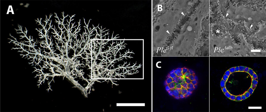
We mainly focus on cytoskeletal linker proteins, in particular plectin, and we study the functional consequences of cytoskeletal organization in cell/tissue mechanics and mechanotransduction, i.e., conversion of physical cues into intracellular mechanosignalling pathways. To fulfil our aims in the complexity of biological systems, we use a combination of in vitro (primary cells and CRISPR/Cas9-targeted cell lines) and in vivo (transgenic models) approaches. Besides conventional molecular biology techniques, we also employ methods that enable us to measure and apply physiologically relevant forces and deformations, such as traction force and atomic force microscopy, magnetic tweezer rheology, cell stretching, and FRET-based tension sensors.
As the team of the Laboratory of Integrative Biology was established as part of BIOCEV (in January 2015), our long-term interest is also defined by BIOCEV Project 1.1.4.: „Mouse models for studying of physiology and pathophysiology of digestive epithelia” (the Functional Genomics Programme). This aim has the following core project objectives: