Institute of Molecular Genetics of the Czech Academy of Sciences
A team of scientists from the Institute of Molecular Genetics of the Czech Academy of Sciences and Charles University led by Vladimír Varga (IMG) managed to optimize the method of expansion microscopy, which has so far been used mainly to study mammalian cells, so that it can now be used to study significantly smaller protozoan cells.
A team of scientists from the Institute of Molecular Genetics of the Czech Academy of Sciences and Charles University led by Vladimír Varga (IMG) managed to optimize the method of expansion microscopy, which has so far been used mainly to study mammalian cells, so that it can now be used to study significantly smaller protozoan cells.
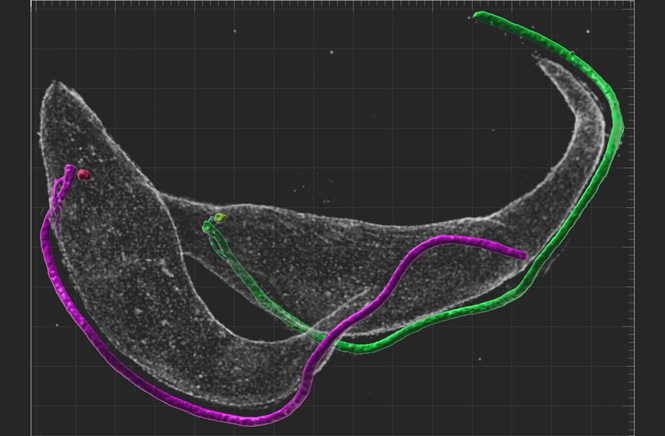
Picture: 3D reconstruction of the cytoskeleton of an expanded cell of Trypanosoma brucei, which is in the final stage of cell division.Source: Vladimír Varga’s team (IMG).
Source: Vladimír Varga’s team (IMG)
Video: Animation of 3D reconstruction of cytoskeleton of expanded Leishmania major cell. Individual structures such as the flagellum and basal bodies are highlighted in color.
Source: Vladimír Varga’s team (IMG)
Gorilak P, Pružincová M, Vachova H, Olšinová M, Schmidt Cernohorska M, Varga V: Expansion microscopy facilitates quantitative super-resolution studies of cytoskeletal structures in kinetoplastid parasites. Open Biol 2021 11(9): 210131. [pubmed] [doi]
Vladimír Varga, Ph.D.,
phone: (+420) 296 443 462,
e-mail: vladimir.varga@img.cas.cz,
web: www.img.cas.cz/vyzkum/vladimir-varga/
Martin Jakubec, Ph.D.,
phone: (+420) 296 443 159, e-mail: m.jakubec@img.cas.cz
Full press release as PDF (in Czech).