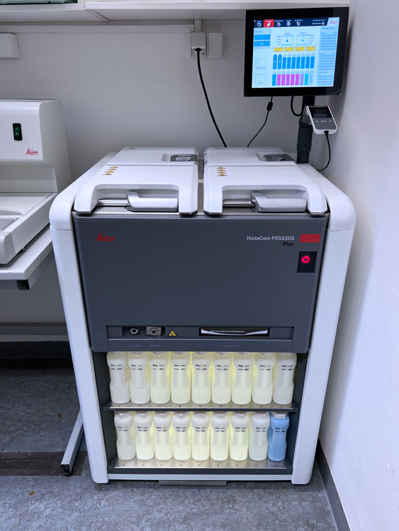
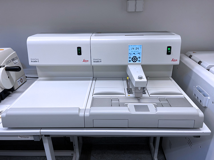
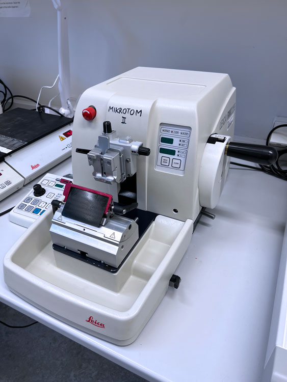
The most important laboratory equipment consists of a set of four Leica devices – tissue processor, paraffin embedding station and two microtomes. There is also a set of trays for tissue deparaffination and pressure cooker for antigen retrieval. Users must be instructed before starting their work on these instruments.
Tissue processor Leica ASP200S is an automated device for tissue dehydration and wax penetration.
Collected samples from registered users are stored in the retort, where they are processed according to the following programme. The programme runs overnight and lasts 16 hours. It is operated by the staff.
Documents | EN manual, CZ manual |
Reservation | reservation form |
Paraffin embedding station Leica EG1150H is the user operated instrument for creating paraffin blocks. The wax-penetrated tissue samples are placed to the heated drawer by the staff. With the help of heated tweezers, users will create the paraffin blocks for tissue sectioning according to their individual preferences. Various sizes of the wax molds are available.
Microtome Leica RM2255
Two Leica RM2255 is a fully motorized rotary microtome equipped with a smooth-running handwheel.
The RM2255 is primarily designed for motorized sectioning, but is also suitable for manual sectioning applications.
The RM2255 allows the sectioning of samples in four different sectioning modes: single stroke, continuous stroke, step mode and rocking mode. The instrument is equipped with an individually adjustable specimen retraction system, which can be switched on and off by the user. The following accessories are available for any of both microtomes:
Trimming thickness range and increments | 1-600 µm |
Section thickness range | 0.5 – 100 µm |
Available feather blade types | A35 – Universal N35 – Hard Tissue R35 – Serial Sections S35 – Routine Sectioning |
Documents | EN manual, CZ manual |
Reservation | Calpendo (“Microtome 1”, “Microtome 2”) |
Vibrating blade microtome Leica VT1200 S
The fully automated Leica VT1200 S vibrating blade microtome is designed for sectioning fixed or unfixed fresh tissue in buffer. Automatic feeding, specimen retraction and the use of a cutting window help to minimize sectioning time. VT1200 S is primarily designed for motorized sectioning, but is also suitable for manual sectioning applications. The vertical deflection of the blade can be measured by the Vibrocheck measurement device. The adjustment of the blade allows minimization of the vertical deflection to below 1 μm, which significantly increases the number of viable cells.
Adjustable amplitude | From 0 – 3 mm, in increments of 0.05 mm |
Frequency | Fixed: 85 Hz (± 10 %) |
Blade travel speed | 0.01 – 1.5 mm/s |
Adjustable cutting window | individually programmable front and rear position |
Maximum specimen size | 33 x 50 x 20 mm |
Total vertical specimen stroke | 20 mm |
Cooling option | Chiller |
Return speed | 1.0 – 5 mm/s, in increments of 0.5 mm/s |
Booking | Calpendo (“Vibratome”) |
Cryostat Leica CM1950
The CM1950 is a high-performance cryostat with an encapsulated microtome and separate specimen cooling. It features a UV disinfection system, an integrated extraction system for section waste and an motor for motorized sectioning. The cryostat is designed to produce frozen sections for biological, medical and industrial applications.
Section thickness range | Setting values 1 to 100 µm 1.0 µm – 5.0 µm in 0.5 µm steps 5.0 µm – 20.0 µm in 1.0 µm steps 20.0 µm – 60.0 µm in 5.0 µm steps 60.0 µm – 100.0 µm in 10.0 µm steps |
Trimming range | 1 – 600 µm (research) 1,0 µm – 10,0 µm in 1,0 µm steps 10,0 µm – 20,0 µm in 2,0 µm steps 20,0 µm – 50,0 µm in 5,0 µm steps 50,0 µm – 100,0 µm in 10,0 µm steps 100,0 µm – 600,0 µm in 50,0 µm steps |
Maximum specimen size | 50 x 80 mm |
Total specimen feed | 25 mm |
Vertical specimen stroke | 59 mm |
Specimen retraction | 20 µm or off |
Specimen orientation | 8° (x-, y-axis), 360° rotation of specimen disc |
Electric coarse feed | Slow: 300 µm/s, in 20 µm steps Fast: 900 µm/s |
Cryochamber | Temperature range: 0°C to -35°C |
Specimen cooling | Temperature range: – 10 to – 50 °C |
Defrosting of specimen head | manual defrost |
Cryochamber defrosting | Automatic cryochamber defrosting: programmable, (hot gas defrost), selectable time, 1 defrost in 24 h or manual hot gas defrost, Defrost time: 12 minutes Automatic shut off defrost: at – 5 °C chamber temperature |
Quick-freeze shelf | Minimum temperature: down to – 42 °C, at chamber temp. – 35°C Number of freezing stations: 15 + 2 Defrost: manual hot-gas defrost |
Peltier element | Number of freezing stations: 2 Maximum temperature difference: 17 K, at chamber temp. of – 35 °C |
Booking | Calpendo (“Cryostat”) |
Trays are compatible with holders for twelve glass slides.
Dewaxing protocol | 3 x 8 min in Xylene 1 x 5 min in Xylene/100% EtOH |
Deparafination is free of charge.
Simple way for the antigen demasking is defined heating in a buffer. The programmable pressure cooker enables defined cooking for 15 or 20 minutes; the entire procedure takes about one hour. Standard 20 mM citrate buffer, pH=6; EDTA pH = 9; or citraconic anhydride buffers are available.
The cooking is free of charge.